Varicose veins and blood flow physics | St. Louis Vein Care
An evidence based guide for St Louis patients
Varicose veins are often explained in one sentence: valves fail, blood falls backward, pressure rises, and veins enlarge. That explanation is directionally useful, but it is incomplete. The venous circulation is not a set of rigid pipes carrying a perfectly uniform liquid under steady conditions. It is a living, branching, highly compliant network whose shape and flow change with standing, walking, breathing, muscle contraction, hydration, temperature, and the pressure outside the vein.
This matters because many medical studies reduce venous disease to measurements that are practical to collect: reflux duration, vein diameter, anatomic location, symptom scores, or whether a vein closes after treatment. Those measurements are valuable. They do not, however, describe every force acting on blood and the vein wall. When a patient’s symptoms or visible vein pattern seems out of proportion to one ultrasound number, the discrepancy may reflect the limits of the measurement rather than a failure of physics.
At Sheen Vein Aesthetics and Functional Medicine in St. Louis, the most useful question is not simply, “Is reflux present?” It is, “How is pressure and volume moving through this individual patient’s venous network, and which abnormal pathway is clinically important?”
The familiar pipe equation and why it is only a starting point
One of the best-known principles of fluid mechanics is the Hagen-Poiseuille relationship. In a long, straight, rigid, circular tube carrying a Newtonian fluid in steady laminar flow, resistance is proportional to viscosity and tube length and inversely proportional to the fourth power of radius. In simplified form, Q = ΔPπr⁴/8μL. Small changes in radius can therefore produce large changes in resistance and flow.
That principle is real, but its assumptions are not fully met in leg veins. Veins deform. Their cross-sections may become oval or partially collapse. Their valves open and close. Varicose segments curve, branch, widen, and narrow. Blood is a suspension of cells and has shear-dependent, non-Newtonian behavior, especially at lower shear rates. Flow is pulsatile and may reverse. Surrounding muscle and fascia apply external pressure. Gravity adds a hydrostatic pressure column when a person stands. A useful equation becomes an incomplete model when its assumptions are treated as the anatomy itself.
The clinical implication is important: a larger vein does not automatically carry proportionally more useful forward flow. Radius tends to lower viscous resistance, but a dilated vein also stores more blood, stretches valve leaflets apart, changes local velocity and shear, and may permit reflux. In a network, flow follows pressure gradients through all available pathways—not simply the largest visible vein.
Gravity creates pressure but walking should interrupt the column
Hydrostatic pressure follows ΔP = ρgh: pressure increases with the vertical height of a fluid column. When a person stands still, gravity therefore raises pressure in dependent leg veins. Competent venous valves divide that column into shorter segments. The calf and foot pumps then compress veins during walking and propel blood toward the heart.
In healthy physiology, ambulation lowers distal venous pressure. In chronic venous disease, reflux, obstruction, impaired calf-pump ejection, reduced ankle motion, or combinations of these factors can prevent pressure from falling normally. This is why ambulatory venous hypertension—not reflux time in isolation—is central to tissue damage, swelling, skin changes, and ulcer risk.
A brief reflux event measured after calf compression is not identical to the pressure history a patient experiences during hours of standing. Duplex ultrasound samples flow at selected locations during a provocation maneuver. It does not continuously measure the total energy losses, pressure transmission, volume displacement, or wall stress throughout the entire network. Air plethysmography and ambulatory venous pressure can add information, but each test also measures only part of the system.
Veins are compliant reservoirs not rigid plumbing
Compliance describes how much volume changes for a given change in pressure. Veins are far more compliant than arteries and hold much of the circulating blood volume. At low pressure, a vein can change shape substantially before it becomes fully round; at higher pressure, further filling stretches the wall and increases wall tension.
Laplace’s relationship provides another useful principle: for a thin-walled cylindrical vessel, circumferential wall tension rises with internal pressure and radius. A dilated vein exposed to sustained pressure therefore experiences greater wall tension. The biology and the mechanics can reinforce each other. Research has identified extracellular-matrix remodeling, altered collagen and elastin, inflammation, smooth-muscle dysfunction, and increased distensibility in varicose veins. As the wall dilates, valve coaptation may worsen; as reflux persists, pressure and wall stress may promote further remodeling.
This feedback loop helps explain why debating whether valve failure or wall failure “came first” can be less useful than mapping the present hemodynamics. The disease is not necessarily a single leak descending from the groin. Reflux may be axial, segmental, tributary-driven, perforator-associated, pelvic in origin, or recurrent through newly connected pathways.
What reflux time can and cannot tell us
Professional guidelines commonly define pathologic reflux using time thresholds, such as more than 0.5 second in many superficial veins and more than 1 second in the common femoral, femoral, and popliteal veins. Thresholds create consistency across laboratories and are essential for diagnosis, research, and coverage decisions.
But a threshold converts continuous physiology into a yes-or-no category. Two patients can both exceed 0.5 second while having very different reflux velocities, volumes, diameters, pressure gradients, symptoms, and collateral anatomy. A vein just below a time threshold is not physically identical to a vein with no reverse flow, and a long reflux time does not by itself reveal how much blood moved backward. Duration, peak velocity, reflux volume, vessel area, and the connected network all influence the hemodynamic consequence.
This is one place where study design may appear not to match physics. The problem is usually not that the study contradicts conservation of mass or fluid mechanics. It is that an easy-to-standardize surrogate—reflux duration—is asked to represent a multidimensional process. Clinical outcomes should therefore be interpreted alongside symptoms, examination, vein mapping, CEAP class, and the patient’s functional goals.
Spider veins make the limitation especially visible
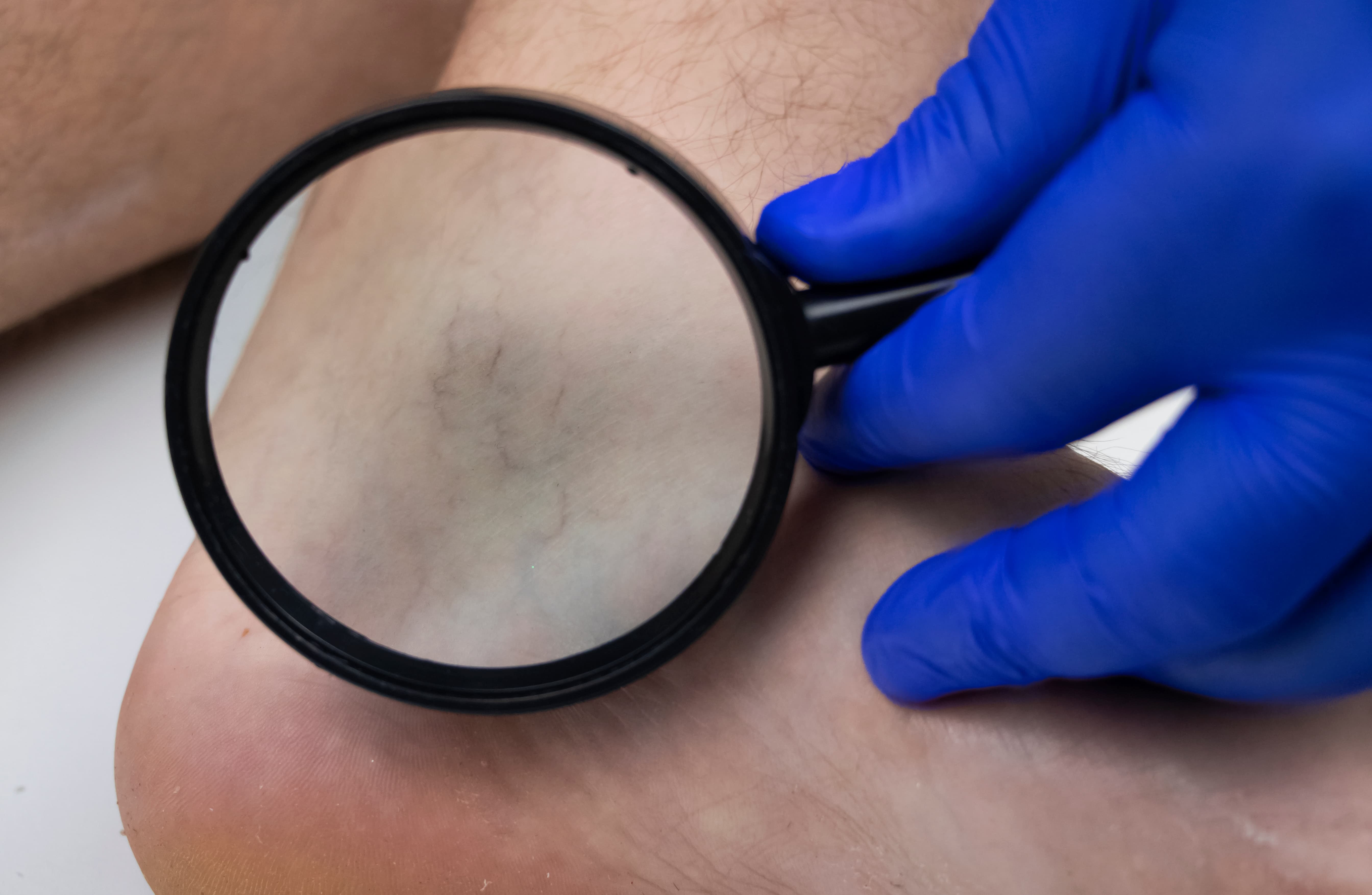
Spider veins, or telangiectasias, are tiny visible intradermal vessels. Reticular veins are somewhat larger subdermal veins and may feed clusters of spider veins. It is tempting to assume that every spider vein must be caused by saphenous reflux. Population evidence does not support that simple rule.
In the Edinburgh Vein Study, increasing telangiectasia severity did not show a clear causal relationship with increasing deep or combined deep-and-superficial incompetence. Other work has found local reticular incompetence near telangiectatic clusters, sometimes with connections to larger superficial or perforating pathways. Both observations can be true: some spider veins are largely local networks, while others coexist with clinically important truncal reflux.
The physics supports this heterogeneity. Pressure is distributed through a network according to pathway resistance, valve behavior, height, boundary pressures, and external tissue forces. A small superficial vessel may enlarge because of local inflow, impaired outflow, wall properties, hormonal and genetic influences, or a feeding reticular vein—even when a standard saphenous segment does not meet a reflux threshold. Conversely, treating only the visible surface cluster without recognizing a significant feeder may increase the chance of incomplete clearance or recurrence.
Why ultrasound technique changes what we observe
Venous duplex is dynamic. Body position, probe pressure, augmentation strength, room temperature, breathing, Valsalva effort, time of day, and the precise sample location can affect diameter and flow. A standing examination generally places the superficial system under the gravitational conditions most likely to reveal reflux. Yet even a well-performed study remains a controlled snapshot.
Modern guidelines appropriately recommend tracing superficial reflux to its source, including saphenous junctions, truncal veins, perforators, tributaries, or pelvic sources. That network-based approach aligns more closely with conservation of mass: blood entering a region must leave it, accumulate temporarily as venous volume, or be redirected through another connection. Closing one path changes the pressure-flow solution for the remaining network.
What this means for varicose vein and spider vein treatment
Good treatment planning begins with the patient rather than an isolated number. Symptoms such as aching, heaviness, swelling, itching, cramping, skin discoloration, or discomfort with prolonged standing should be evaluated in context. Visible varicose veins may warrant a comprehensive reflux ultrasound. Spider veins without symptoms or larger abnormal veins may be primarily cosmetic, but a broader evaluation can be appropriate when the distribution, recurrence, or symptoms suggest an underlying source.
Treatment may include compression, walking and calf-pump conditioning, elevation, weight management when relevant, ultrasound-guided or visual sclerotherapy, non-compounded foam treatment, or endovenous laser ablation. The right sequence depends on anatomy and clinical need. Treating significant upstream or feeding reflux before surface veins often makes hemodynamic sense, but not every spider-vein patient has truncal disease and not every detected reflux segment requires intervention.
The purpose of closing an incompetent superficial vein is not to “block circulation.” It is to remove an inefficient pathway and redirect blood through competent superficial and deep channels. Because the circulation is a network, the result depends on adequate deep venous outflow, collateral capacity, muscle-pump function, and the accuracy of the original map.
A better way to connect studies with physics
Clinical vein research becomes more physically complete when it combines anatomy, reflux duration, velocity or volume, pressure-related measures, vessel compliance, muscle-pump function, posture, and patient-centered outcomes. New fluid-structure interaction models are beginning to represent flexible valve leaflets and deformable vein walls, but models still simplify real biology and require in-vivo validation.
For patients, the practical message is straightforward: one diameter or reflux time is useful, but it is not the whole diagnosis. For clinicians and researchers, the opportunity is to measure the venous system more like the living pressure-flow network it is. Physics does not invalidate evidence-based vein care. It explains why individualized mapping and clinical judgment remain necessary.
Schedule a vein evaluation in St Louis
If you have painful varicose veins, recurrent spider veins, leg heaviness, swelling, skin changes, or symptoms that worsen with standing, Sheen Vein Aesthetics and Functional Medicine offers physician-guided evaluation and personalized treatment planning in South St. Louis County. Medical vein care may be insurance-based when coverage criteria are met; spider-vein treatment is generally cosmetic.
Call the office to schedule a consultation at 11144 Tesson Ferry Road, Suite 100, St. Louis, Missouri 63123. An examination and, when indicated, a properly performed venous duplex ultrasound can help determine whether your concern is local, cosmetic, or part of a larger reflux pattern.
Frequently Asked Questions
Can spider veins exist without varicose veins or saphenous reflux?
Yes. Spider veins frequently occur without measurable saphenous insufficiency. Some are associated with local reticular or feeding veins, while others coexist with larger reflux pathways.
Does a larger vein always mean worse venous disease?
No. Diameter can correlate with reflux, but it does not independently define symptoms, pressure, reflux volume, or treatment need. Anatomy and function must be interpreted together.
Why is a standing venous ultrasound important?
Standing increases the gravitational pressure column and helps reveal abnormal reverse flow that may be less apparent when a patient is lying down.
Does closing a varicose vein reduce circulation?
When the deep system is patent and treatment is appropriately planned, closing an incompetent superficial vein redirects blood toward healthier return pathways rather than depriving the leg of circulation.
Educational disclaimer: This article is for general education and does not diagnose a condition or replace an individual medical evaluation. Treatment recommendations and insurance coverage depend on symptoms, examination findings, ultrasound results, medical history, and plan requirements.
References
3. Secomb TW. Hemodynamics. Comprehensive Physiology. 2016.
6. Ruckley CV, et al. Telangiectasia and venous reflux in the Edinburgh Vein Study. Phlebology. 2012.